3D Imaging
Jupiter, FL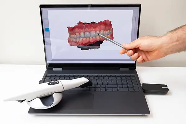
Wisdom teeth, or third molars, are among the most variable teeth in the mouth, both in how they develop and in how they position themselves relative to nerves, sinuses, and adjacent teeth. At Roy C Blake, III DDS, MSD Maxillofacial Prosthodontist, three-dimensional imaging gives our team a precise understanding of this anatomy before any extraction is recommended, supporting safer, more predictable treatment.
Why Wisdom Teeth Require Careful Evaluation
Most people develop four wisdom teeth, typically emerging in the late teenage years or early twenties. Because they are the last teeth to develop, they often do not have enough room to erupt fully or in proper alignment. This can result in teeth that are impacted, meaning they remain partially or fully trapped beneath the gum and bone, sometimes growing at an angle that affects neighboring teeth.
The challenge with wisdom teeth is that their position varies significantly from patient to patient, and even from one side of the mouth to the other. A tooth that appears straightforward on a standard X-ray may actually be close to a major nerve or sinus cavity, details that are easy to miss on a flat, two-dimensional image. Without a clear understanding of this anatomy, planning a safe extraction becomes far more difficult.
What 3D Imaging Reveals Before Extraction
Cone beam computed tomography provides a complete three-dimensional view of each wisdom tooth and the structures surrounding it. This allows our team to evaluate the case with a level of detail that significantly improves both safety and predictability.
Specific findings that 3D imaging makes visible include:
| • |
The exact position and angulation of each wisdom tooth relative to neighboring teeth
|
| • |
The proximity of lower wisdom tooth roots to the inferior alveolar nerve, which controls sensation in the lower lip and chin
|
| • |
The relationship between upper wisdom teeth and the maxillary sinus
|
| • |
Root shape and curvature, which affects how a tooth will be removed
|
| • |
The density and quality of surrounding bone
|
| • |
Any cysts or other abnormalities associated with an impacted tooth |
This information allows our team to anticipate challenges before treatment begins rather than discovering them mid-procedure, contributing to a smoother, more predictable extraction experience.
Reducing the Risk of Nerve Involvement
One of the most important reasons for 3D imaging before wisdom tooth extraction is evaluating the relationship between the lower wisdom teeth and the inferior alveolar nerve. This nerve runs through the lower jawbone and is responsible for sensation in the lower lip, chin, and portions of the gum tissue. In some patients, the roots of the lower wisdom teeth sit very close to this nerve, or in certain cases, the nerve canal even passes directly between the roots.
A standard panoramic X-ray can suggest a wisdom tooth root and the nerve canal are close together, but it cannot show their true three-dimensional relationship. A 3D scan removes this ambiguity, allowing our team to see precisely how close the roots come to the nerve and from which direction, directly informing the surgical approach and helping reduce the risk of nerve-related complications.
Determining the Right Time for Extraction
Timing plays a significant role in wisdom tooth treatment planning, and 3D imaging helps our team evaluate not just whether extraction is needed, but when it makes the most sense. In younger patients, wisdom tooth roots are often still developing, which generally makes extraction more straightforward and recovery more predictable. As roots mature and become more fully formed, they tend to sit closer to the nerve canal and other surrounding structures, which can make later extractions more complex.
By using 3D imaging to evaluate root development and position at an earlier stage, our team can identify patients who may benefit from proactive extraction before complications develop. This is particularly relevant for wisdom teeth that show early signs of crowding, abnormal angulation, or limited space for proper eruption, since these patterns often do not improve with time.
Identifying Teeth That May Not Need Extraction
Not every wisdom tooth requires removal. In some cases, a wisdom tooth is positioned well, has adequate room to function, and shows no signs of impaction or disease. Three-dimensional imaging helps our team make this determination with confidence rather than recommending extraction as a default precaution. When a tooth is fully erupted, properly aligned, and free of decay or gum disease, our team may recommend monitoring the tooth over time rather than proceeding with removal.
This careful, individualized evaluation reflects a broader commitment to recommending treatment based on what each patient's anatomy actually requires, rather than applying the same approach to every case.
Planning for Upper Wisdom Teeth and the Sinus
Upper wisdom teeth present a different anatomical consideration, since their roots are often positioned close to the maxillary sinus. Removing a tooth with roots that extend into or near the sinus floor requires careful planning to avoid creating an unwanted communication between the mouth and sinus cavity. Three-dimensional imaging allows our team to clearly see this relationship beforehand, informing both the surgical technique and any precautions needed during healing.
Supporting an Informed Decision
Beyond improving surgical safety, 3D imaging also helps patients understand their own case more clearly. Rather than relying on a verbal description of risk, our team can show you the exact position of your wisdom teeth relative to the nerve, sinus, or adjacent teeth using the same images used for treatment planning. This visual understanding often makes it easier for patients to feel confident in the recommended approach, whether that means proceeding with extraction at our practice or, in more complex cases, referral to an oral surgeon.
When to Consider Wisdom Tooth Imaging
Our team typically recommends 3D imaging when a wisdom tooth appears impacted, when its position on a routine X-ray suggests proximity to the nerve or sinus, or when a patient reports pain, swelling, or difficulty opening the mouth fully. Younger patients are sometimes evaluated proactively, since root development is often less advanced and extraction tends to be more straightforward earlier rather than later.
Schedule Your Evaluation
If you or a family member has been told that wisdom tooth extraction may be needed, Roy C. Blake III, DDS, MSD, Maxillofacial Prosthodontist can provide the detailed imaging necessary to plan treatment with confidence. Call us today at (561) 296-3399 to schedule an evaluation and learn more about what 3D imaging can reveal about your specific case. |